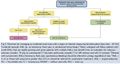
Uncategorized files
Jump to navigation
Jump to search
Showing below up to 142 results in range #1 to #142.
 2019AdnexalCyst.pdf ; 1.14 MB
 4ac4f1dfa9f5e Territory.jpg 500 × 980; 156 KB
 AAA.png 1,158 × 902; 412 KB
 Acradrenal1.JPG 740 × 860; 99 KB
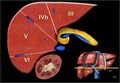
 Acrpanc1.JPG 925 × 753; 128 KB
 Acrpanc2.JPG 921 × 860; 132 KB
 Acrpanc3.JPG 911 × 743; 119 KB
 Acrpanc4.JPG 923 × 749; 109 KB
 Acrpanc5.JPG 926 × 755; 124 KB
- Acrpancfull.pdf ; 2.68 MB
 Acrrenal1.JPG 1,384 × 779; 195 KB
 Acrrenal2.JPG 1,380 × 792; 180 KB
 Acrrenal3.JPG 1,375 × 739; 138 KB
 Acrrenal4.JPG 920 × 825; 170 KB
 Acrrenal5.JPG 917 × 490; 91 KB
 Acrrenal6.JPG 923 × 274; 66 KB
 Adnexa.png 1,010 × 1,266; 372 KB
 AdnexaTable.png 1,316 × 730; 380 KB
 Adnexal2019-1.jpg 2,167 × 1,070; 299 KB
 Adnexal2019-2.jpg 2,178 × 1,071; 303 KB
 Adnexal2019-3.jpg 1,800 × 1,240; 366 KB
- Adnexalmass.pdf ; 672 KB
 Adnexalmass1.jpg 964 × 1,030; 187 KB
 Adnexalmass2.jpg 983 × 394; 77 KB
 AdrenalMRIquant.jpeg 756 × 199; 52 KB
 Aspects.jpg 300 × 302; 34 KB
 Atlanta2.jpg 961 × 493; 109 KB
 Atlanta 1 from Radiographics.jpg 985 × 540; 75 KB
 BIRADS Card.JPG 2,592 × 1,936; 1.93 MB
 Body MRI Cheat Sheet v3.docx ; 26 KB
 Bozniak.JPG 921 × 387; 101 KB
 Bozniak 2019.jpg 835 × 792; 258 KB
- CT Perfusion Billing Rules.pdf ; 133 KB
 Ctperfusion.jpg 702 × 211; 28 KB
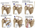 DC-Joint-Classification.png 632 × 501; 422 KB
 Dens.png 600 × 412; 32 KB
 ECUNormalSignal.png 758 × 1,084; 596 KB
- FirstTrimesterViability.pdf ; 127 KB
 Fleischner 2017.jpg 832 × 839; 152 KB
- Fleishner IPF Guidelines2018.pdf ; 7.86 MB
- Flesichner Guidelines 2017.pdf ; 1.89 MB
- Fluoro Contrast May11.xlsx ; 12 KB
 GBBileDuct.png 1,784 × 958; 950 KB
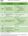
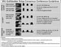 GB Polyp Guidelines.jpg 1,094 × 852; 183 KB
- GB Polyp PDF.pdf ; 2.15 MB
 Incidental Thyroid Nodule.jpg 1,346 × 929; 226 KB
- JPAD ARIA.pdf ; 729 KB
- LIRADS 2017 Core.pdf ; 703 KB
 LIRADS 2017 img1.jpg 682 × 838; 104 KB
 LIRADS 2017 img2.jpg 697 × 876; 128 KB
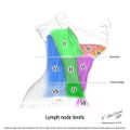 LN levels.jpg 630 × 630; 142 KB
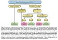
 LiverIncidental.png 1,371 × 632; 324 KB
 LiverIncidental2.png 1,484 × 635; 160 KB
 Liver Segment Lower.jpg 440 × 306; 20 KB
 Liver Segment Upper.jpg 440 × 293; 17 KB
- Liver doppler.pdf ; 20.11 MB
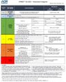
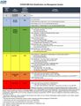
- LungRADSAssessmentCategoriesv1-1.pdf ; 657 KB
- MRI Cheat Sheet.docx ; 17 KB
 O-RADS Table1.jpg 1,039 × 1,282; 420 KB
 O-RADS Table2.jpg 1,027 × 1,305; 318 KB
- O-RADS US Lexicon Key Terms.pdf ; 454 KB
- OBUS Viability Article.pdf ; 1.4 MB
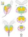
 OB anomalies.JPG 1,577 × 770; 261 KB
 ORADS MRI.jpg 930 × 1,208; 315 KB
 Optn.gif 1,038 × 379; 105 KB
- PIRADS V2-1.pdf ; 2.84 MB
- PIRADS V2.pdf ; 2.45 MB
- Perianalfistula1.pdf ; 5.49 MB
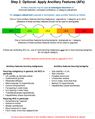
 Pirads-dwi.png 717 × 332; 68 KB
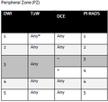 Pirads PZ.png 276 × 259; 6 KB
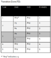 Pirads TZ.png 404 × 457; 21 KB
 Pirads dce.png 499 × 120; 9 KB
 Pirads t2.png 506 × 575; 83 KB
 Pirads zones.png 457 × 610; 236 KB
- RG ARIA.pdf ; 6.36 MB
- RVU Table v3.xlsx ; 12 KB
- RVU Table v4.xlsx ; 12 KB
- RVU Table v5.xlsx ; 12 KB
 Rad-icon.png 125 × 125; 17 KB
- Radiographics Atlanta Pancreatitis.pdf ; 14.59 MB
 Rectal Ca staging.png 1,914 × 1,030; 431 KB
- Rectalcancer.pdf ; 4.5 MB
- Residents.pdf ; 9.41 MB
- Rgadrenalmri.pdf ; 3.74 MB
- SOG BreastUSRev02-14-2017.pdf ; 498 KB
- SOG Diagnostic Breast MRI.doc ; 98 KB
- SOG MRI-GuidedBreastBiopsy.doc ; 71 KB
- SOG Outreach.doc ; 72 KB
- SOG Outreach ACRattachment.pdf ; 205 KB
- SOG USGuidedCoreBiopsy.doc ; 50 KB
 SVT guideline.JPG 801 × 1,122; 193 KB
 Selection 061.png 249 × 194; 14 KB
- Spine Level Labeling.pdf ; 409 KB
 Spleensize.png 759 × 854; 100 KB
- StrokeCT.pdf ; 2.1 MB
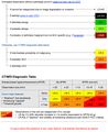
- TI-RADS JACR 2017.pdf ; 556 KB
 TIRADS.jpg 1,342 × 1,012; 244 KB
- Turbo Presentation.pptx ; 2.06 MB
 UIP Criteria.jpg 1,350 × 524; 188 KB
- UIP Criteria Full PDF.pdf ; 1.73 MB
- Umbilical artery Doppler.pdf ; 4.18 MB


